Equine Sports Medicine and Rehabilitation Services
Our equine sports medicine and rehabilitation services are intended to treat soreness and injuries, joint issues and inflamation.
Cold Compression Therapy
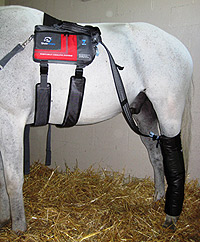
Cold water has long been used as an anti-inflammatory and analgesic for all animals. Combining dry cold temperatures with alternating pressure greatly improves the removal of heat and edema while improving circulation. The circulating cold water removes heat and reintroduces colder water every few minutes.
The alternating compression drives the cold deep into the tissues then releases it allowing for improved circulation and further reducing inflammation. Cold compression therapy has been successfully used for horses with tendon injuries, sore muscles, cellulitis/lymphangitis or even as a post operative aid to reduce swelling and pain.
Extracorporeal Shockwave Therapy
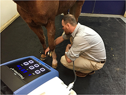
The Equine Specialty Hospital uses extracorporeal shockwave therapy as a form of healing in horses. Extracorporeal shockwave therapy is a noninvasive modality (extracorporeal means "outside the body") that uses sound waves to stimulate healing in wounds, ligaments, tendons and bony structures. Horses are usually sedated for the therapy session and treatments last no longer than 30 minutes.
Extracorporeal shockwave therapy increases blood flow, increases growth of new blood vessels and increases the production of natural healing factors in the treated area which all result in improved speed and quality of tendon, ligament, bone, and wound healing. It is often used in cases of suspensory ligament injury, tendon disease, navicular syndrome, neck or back pain and injuries, bone bruising, bucked shins and many other orthopedic problems. It also provides mild, short-term pain relief.
Stem Cell Therapy
Stem cell therapy is used for the treatment of tendon, ligament, and joint diseases in the horse. A stem cell has pluripotency (it can become any type of cell) or multipotency (it can become any cell, but with limitations). Technically stem cells can be obtained from any tissue; however, bone marrow-derived has the most scientific evidence supporting efficacy. We collect bone marrow and, under special laboratory conditions, stem cells can be cultured. These can be placed in areas of injury, where they can integrate into local tissue and aid in repairing injury/reducing inflammation.
Equine Stem Cells, enables us to take cells from a horse and implant the cultured tissue back into the same horse (known as autologous therapy). We never use cells from one horse to treat another. Stem cells are usually for a tendon injury or intra-articular soft tissue injury. Implantation of the stem cells is a straightforward procedure, performed under sedation with or without an ultrasound to guide the injection. This can be performed on an out-patient basis. The veterinary surgeon responsible for your horse will discuss the potential benefits and risks associated with stem cell therapy, together with post-implantation care and exercise. Complications are very rare. In most cases, we like to see the horse for follow-up assessment one month after injection.
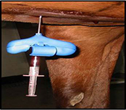
Platelet Rich Plasma (PRP)
Historically soft tissue injuries are very slow to heal because tendons and ligaments have a poor internal blood supply. Without a good blood supply to bring in the growth factors required for healing, it takes a long time for the injury to heal and it heals with poorly organized, weak scar tissue. The growth factors are produced and stored in platelets, which are harvested and injected as Platelet Rich Plasma. Injecting PRP directly into the injury greatly improves the speed of healing and the scar tissue is stronger and more organized.
Superficial and deep digital flexor tendonitis (bowed tendons), suspensory desmitis, and many other tendon and ligament injuries are candidates for PRP therapy. The location and severity of the injury determine if and how PRP is utilized.
Ultrasound examination of the injured limb, performed in combination with a detailed lameness evaluation, identifies the location and severity of the injury within the specific tendon or ligament. If the injured portion of the tendon or ligament is surrounded by a tendon sheath, PRP is injected directly into the tendon sheath. If the injury is located outside of a tendon sheath, the ultrasound examination must be able to demonstrate a “core lesion” or distinct hole, in the tendon or ligament. PRP is then administered directly into the core lesion via ultrasound-guided injections.
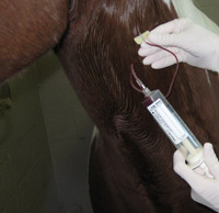
PRP is acquired by first obtaining a sample of your horse’s own blood. The sample is then inserted into a special centrifuge tube and centrifuged for 15 minutes. This process separates the sample into “Platelet Rich Plasma,” red and white blood cells, and non-platelet rich plasma. A stopcock attached to the special centrifuge tube allows fast and easy harvesting of PRP. Following sterile preparation of the injury site, the PRP is injected into either the tendon sheath or core lesion. Your horse does not require hospitalization for PRP therapy.
Interleukin 1-Receptor Antagonist Protein (IRAP)
IRAP is available for treating arthritis, synovitis, and cartilage damage within joints. This anti-inflammatory protein is harvested from the horse’s own blood and works by stopping the production of Interleukin 1 within the joint.
Interleukin 1 is a protein produced by the joint that causes inflammation, which in turn leads to joint damage. IRAP binds to the site of Interleukin 1 production within the joint, effectively preventing release of this damaging protein.
IRAP is harvested by first drawing a blood sample into a syringe containing glass beads coated with chromium. Chromium stimulates the production of IRAP during a 24 hour incubation period at 37 C. After the sample has incubated for 24 hours, the sample is centrifuged and the IRAP is collected. Up to 4 doses can be obtained from a single syringe and the extra doses are frozen and stored for later use.
IRAP is much less irritating to the joint’s cartilage than steroids and helps prolong joint health. It is injected into the joint every 3 weeks for a total of 3-4 doses in lieu of steroid & hyaluronic acid injections. The series of IRAP injections can be repeated at any time.